Migrating border cells labeled with green (membrane-GFP) and red (F-tractin). Border cells move as a group to the oocyte at right. Movie by Yujun Chen and Jocelyn McDonald.
Border cells (red) use non-muscle myosin II (myo-II; green) to migrate through the nurse cells. Movie by Yujun Chen and Jocelyn McDonald.
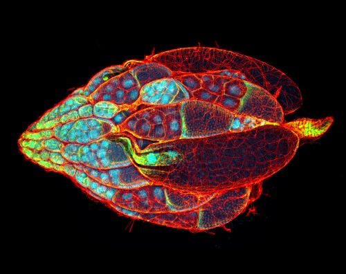
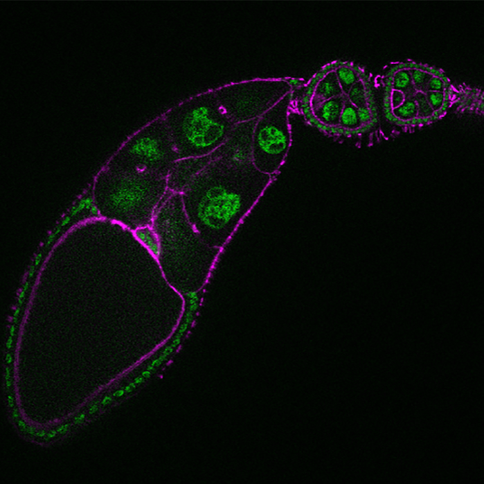
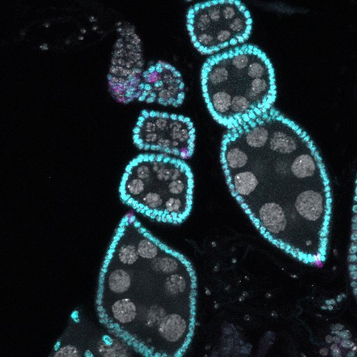
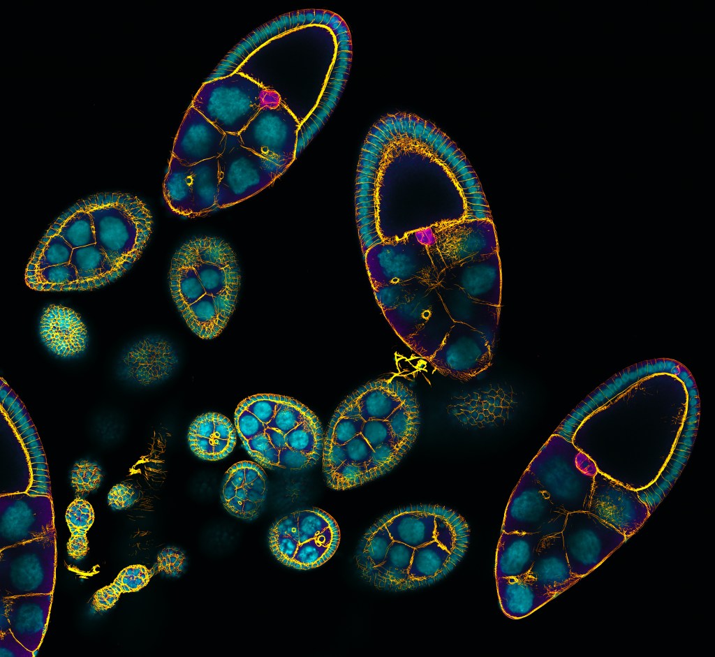
Migrating border cells labeled for a cell membrane marker (PLC∂PH-GFP). Dynamic cellular behaviors, including protrusions, cell movement, and cell shapes can be seen. Movie by Yujun Chen.A whole ovary labeled for nuclei (cyan) and F-actin (red). Image by Yujun Chen.An ovariole stained to visualize nuclei (green) and cells (magenta). Border cells are at the oocyte in the oldest egg chamber. Image by Nirupama Kotian.Ovarioles stained for germline nuclei in gray, somatic nuclei in cyan, and polar cells in purple. Image by Luke MesserEgg chambers stained for F-actin (gold) and nuclei (cyan) and a marker for border cells (pink). Image by Yujun Chen.Ovarioles stained for Singed (Fascin, red), nuclear Lamin (gold) and DAPI (blue). Image by Yujun Chen.